Harvard Researchers Validate Metalens-Based QPI Endoscopy Using Phasics SID4-HR
Why phase matters in endoscopy
In biomedical imaging, many optical techniques are capable of visualizing semi-transparent or weakly scattering tissues, but they often rely on exogenous contrast agents such as fluorescent dyes or labels. Quantitative Phase Imaging (QPI) offers a powerful alternative by providing intrinsic, label-free contrast that reflects variations in refractive index and thickness within the sample. This makes QPI particularly effective for thin and weakly absorbing specimens, where traditional amplitude-based microscopy fails to yield sufficient structural information.
Endoscopy, by contrast, is designed for in vivo imaging of thick, highly scattering tissues, where light propagation becomes dominated by multiple scattering and absorption. Under these conditions, recovering quantitative phase information is considerably more challenging. Nevertheless, recent advances have demonstrated that QPI principles can be integrated into endoscopic systems through innovative optical and computational approaches.
Although imaging thick and diffusive tissues remains a persistent challenge, not only for QPI but for biomedical imaging in general, combining phase-based contrast with endoscopy represents a promising direction toward quantitative, minimally invasive optical diagnostics.
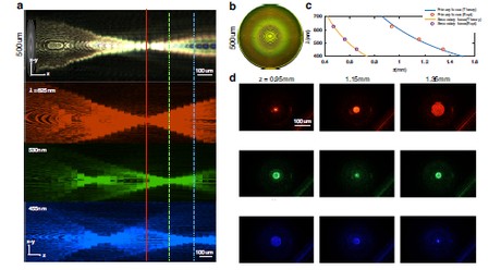
A recent study from Harvard University and Massachusetts General Hospital, published in Light: Science & Applications, demonstrates a compact, interference-free route to quantitative phase imaging intrinsically compatible with endoscopy. The approach leverages a metalens whose focal length shifts with wavelength, under broadband illumination, a color camera’s RGB channels correspond to distinct defocus planes along the optical axis, an effect known as chromatic focal shift.
By integrating this multi-plane information within the Transport of Intensity Equation (TIE) framework, the researchers reconstructed quantitative phase maps directly from a single color image, without any interferometer or mechanical scanning.
Unlike conventional multi-frame or interferometric QPI methods, this strategy retrieves the phase from a single color exposure. It removes the need for additional reference optics or mechanical movement, simplifying the optical design while preserving the quantitative nature of phase imaging.
Calibration and validation with PHASICS: from test targets to biological samples
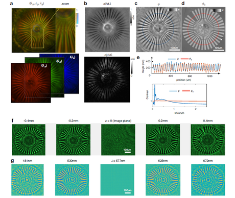
Before applying the technique to living specimens, the researchers validated its performance using a Siemens star phase target. This allowed them to test the system under well-defined conditions: they computed the contrast-transfer function (CTF) to quantify spatial resolution, and compared the reconstructed phase with a reliable reference to assess accuracy.
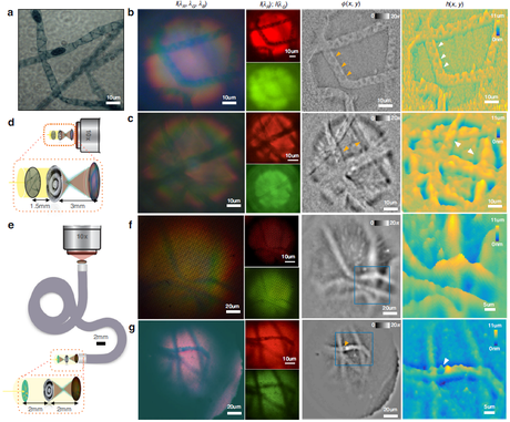
For the reference, they used a Phasics SID4-HR quantitative phase imaging camera, chosen for its high sensitivity, stability, and well-established quantitative precision. The SID4-HR has been widely adopted by research groups worldwide as a ground-truth instrument for phase measurements, making it an ideal benchmark for this study.
In practice, the SID4-HR was attached to the port of a conventional transmission microscope to record the same Siemens target. The resulting reference phase map was then compared point by point with the reconstruction obtained using the metalens method.
The comparison confirmed that the new approach retrieves height variations with an accuracy of approximately 0.1 λ and achieves a lateral resolution close to 1 μm, sufficient to capture cell-level structures.
To further test real-world feasibility, the team mounted the metalens directly at the tip of a coherent fiber bundle (CFB). In typical conditions, fiber bundles disrupt spatial phase coherence, making QPI extremely difficult. In this configuration, however, the phase information was pre-encoded into intensity through the chromatic defocus among RGB channels. As a result, the phase could still be faithfully recovered after transmission through the fiber bundle.
Under these conditions, the team imaged Spirogyra samples and clearly visualized subcellular structures such as spiral chloroplast bands and the relative depth of overlapping filaments. The experiment demonstrated that the method remains effective in realistic endoscopic scenarios.
Assessing the reliability of PHASICS quantitative measurements
Whenever a new imaging technique is introduced, a fundamental question arises: Can the quantitative results be trusted?
This is a question faced by nearly every development team:
- Is the reconstructed phase truly accurate?
- How can I prove that the results are reliable?
- Is there an established instrument that can serve as a validation reference?
In this work, the researchers’ decision to include a recognized quantitative reference provides that assurance. By comparing their new approach with an independently validated system, they avoided the pitfall of self-validation and reinforced the scientific credibility of their results.
Here, the Phasics SID4-HR served as the trusted benchmark. It was not meant to replace the new technique, but to anchor it to a known quantitative standard, enabling the findings to be confidently accepted by the broader research community.
We sincerely thank the Harvard and Massachusetts General Hospital team for their outstanding contribution to advancing quantitative phase imaging and for trusting Phasics technology as a reliable reference in their work.
At Phasics, we continue to support the research community with quantitative imaging tools that enable next-generation optical diagnostics.
Contenu connexe


Jun 29th 2026
Exosens conclut un contrat avec BROLIS
Exosens conclut un contrat majeur de long terme avec BROLIS pour fournir 17 000 tubes intensificateurs de lumière aux forces armées tchèques

Jun 26th 2026
Visit our booth #514 during OPTIFAB

Jun 25th 2026
Innover pour la défense et la sécurité
Exosens obtient un financement de 140 millions d’euros de la BEI pour soutenir l’innovation dans l’industrie Européenne de la défense et de la sécurité

Jun 22nd 2026
When Cells Stop Dividing, Do They Stop Growing?

Santa Cruz, California.
DE Aug 22nd 2026 A Aug 28th 2026
Aeroballistic Range Association 2026
Retrouvez Exosens lors de la conférence ARA, du 23 au 28 août, à Santa Cruz, en Californie.