Among the investigative techniques applied to art inspection and restoration, infrared imaging plays an important – and rapidly growing – role. By deploying appropriate sensors and cameras with high resolution art restorers and conservators can effectively engage in the examination of valuable art works and their preservation as a cultural heritage.
Cultural Heritage
For many centuries right up to the present artists have applied paint to canvas, wood, parchment, paper, ceramics and walls to create images of their own intuition and to portray their contemporaries. They have left works of the highest value that should endure the test of times. However, like all works created by humans, paintings are damaged by exposure to sunlight, adverse environments, chemical reactions and other forces of nature, which attack their substance and appearance. Restoration and conservation is meant to slow this process of decay, stop or reverse it.
Even in economically difficult times preservation of a cultural heritage for future generations counts among the important societal responsibilities. Museums and independent art research institutions are called to this task, such as IPARC (International Platform for Art Research and Conservation) in Belgium, which employs a multi-disciplinary team offering restoration and conservation services to collectors and the general public alike.
Key to professional preservation is exact knowledge of the physical and chemical composition of a work to be preserved; besides information what forces have contributed to its present material state. However, the integrity of the work must not be changed or diminished by the analytic methods deployed. In other words, only non-invasive examination techniques should be used.
Broad Range of Technologies
Radiation – if used at moderate strength and with limited exposure to the work – is considered a more or less non-invasive tool. Therefore, radiation tools have become ubiquitous in the examination and preservation of cultural goods. Radiation sources in the xray realm take first place: an x-ray fluorescence analysis can uncover the alloys often used in the metal foils of late medieval paintings. Classical x-ray examination also penetrates multi-layered painted panels to understand their structural principles. A subsequent ultraviolet fluorescence analysis can then determine the surface properties such as lacquer covering, retouching and rework. Based on such localization procedures more in-depth analysis procedures can be initiated.
The spiritual message of a painting lies in its wholesome appearance in the visible realm since this impression is immediately accessible to the human eye. A conservator will go beyond this appearance. He or she will look at the work under direct incident light as well as oblique light at a rather flat angle to determine an artist’s intentions and, through the use of the proper analytical tools, to distinguish between its original state and later additions and damages.
Almost the entire frequency spectrum from the visible realm to the Terahertz range can be used for art examination purposes (Figure 1). Penetration is deeper at lower frequencies. Therefore procedures that intend to explore hidden layers are usually located in the infrared realm. A look beneath the surface of a painting is of special interest because it will deliver information about the history of its creation, its structure and, specifically, about underpaintings or underdrawings.
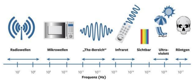
Figure 1. Almost the entire frequency spectrum can be used for art examination purposes. Source: Stern online Infografik.
Relatively new to the field of art examination and conservation is the use of frequencies in the Terahertz realm, next to the infrared.
Short Wave Infrared for Deep Penetration
Of the many art investigation procedures based on short-wave infrared radiation, two typical procedures shall be briefly introduced: Infrared reflectography and optical coherence tomography (OCT).
Both methods employ the idea that most artist paints are more or less transparent to short-wave infrared radiation and therefore allow a look at underlying patterns and structures. As an example serves Figure 2 [1]. It is a painting panel depicting the “Holy Ursula and Eleven Thousand Virgins” by the Flemish painter Pieter Claeissens I, on display at the Museo de Bellas Artes de Oviedo Asturias, Spain.
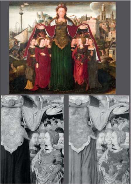
Figure 2. Panel painting “Holy Ursula and Eleven Thousand Virgins” by the Flemish artist Pieter Claeissens I, captured in the visible spectrum (top), a section of it in the NIR realm 0.9 to 1.0 μm (lower left), and in the short-wave infrared (SWIR) from 0.9 to 1.7 μm wavelength. Source: [1].
A closer look at the work shows that there is no obvious pattern visible in the left-hand side of the Saint’s green garment. Also, the part in the lower left exhibits no visible drapery. The picture was taken with an NIR (near infrared) camera, whose sensitivity is limited, due to the image converter based on silicon, to the realm around 1 μm. Therefore the green color is not sufficiently transparent to it. On the other hand, a camera equipped with an image sensor in InGaAs technology and a spectral sensitivity covering wavelengths from 0.9 to 1.7 μm will deliver the image in the lower right of Figure 2. It clearly shows the hidden green folds in the garment.
The method of IR reflectography is frequently used to uncover a painting’s underdrawings. They are not really meant to be seen by the regular observer since the artist had them covered and hidden by the actual visible patterns. However, in an art historical view there is great interest in these underdrawings because they lead to a deeper understanding of the artist’s original intention and creation processes.
An SWIR analysis of a painting does not always deliver satisfactory results. Its success strongly depends on the materials of the underpainting and the layers on top of it. There must be a sufficient optical contrast to distinguish between these interwoven patterns. Underdrawings carried out with charcoal pen and graphite on a light ground will be well recognizable. Red crayon on a white surface or white crayon on dark ground are barely visible under IR radiation. A layer of paint on top of an underdrawing will not block the infrared illumination, especially with thin application and light colors at the red end of the visible spectrum, such as skin tones in portraits.
However, heavily applied layers of paint and high carbon content in their pigment will absorb much of the radiation energy so that an analysis using the entire SWIR realm is not possible since absorption may strongly vary with frequency. Contemporary examination techniques therefore tend to use multiple narrow-band spectral areas. This involves taking digital photographs in the visible realm and at various SWIR wavelengths. They are overlaid in a computer program and processed by appropriate algorithms to a degree that finally reveals any existing underdrawing.
A second method, which is more and more deployed, is optical coherence tomography. It is widely and successfully used as a medical diagnosis tool. Lately, OCT is gaining importance also in the field of art examination since it enables cross-sectional views of the objects under test without touching or damaging them.
The principle of a full-image OCT is explained in Figure 3. It is based on the interference of a direct light beam with a second beam that is reflected from the object under test. Both of them originate from the same light source but have been split by a semitransparent mirror or beam splitter before being reunited. A computational combination of several of such interference images taken by an SWIR camera will compose the final tomographic image. Basically this is an interference microscope, where the object under investigation is illuminated by a white light source, such as a halogen lamp.
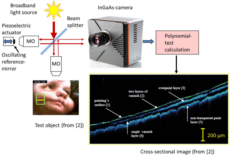
Figure 3. OCT setup and tomogram in the SWIR realm of a touch-up applied on the cheek of the child in the painting “Madonna dei Fusi“ by Leonardo da Vinci. Source: Tomogram in [2].
Figure 3 shows a layout resembling a Michelson interferometer with identical lenses (MO) in both arms. The 2D SWIR camera captures a series of interference images which differ only in their phase distribution. Phase shift is induced by slight displacements of the reference mirror at a fraction of the light wavelength via a piezo electric micro-actuator. Combining these interference images through a higher-order polynomial establishes the tomographic image – a cross sectional view of the object under test.
Using this OCT test procedure a work by Leonard da Vinci, “Madonna die Fusi“, was examined in an area that under UV illumination exhibited a somewhat odd appearance: an irregularity on the right cheek of the child [2]. OCT analysis tracing the yellow line in the green rectangle uncovered the layer sequence shown in the lower right of Figure 3. At the very bottom there is an intransparent layer of paint (5). On top of this is the varnish (4). Surprisingly, there appears a second color layer (3) above the varnish, which usually is applied by the artist at the end. It obviously stems from a retouching carried out afterwards. The entire ensemble was then professionally covered by another layer of varnish (2).
Since the reflected signals originate from the depth of the object they are heavily attenuated. Thus the examination requires an SWIR camera with wide dynamic range and low noise. The cameras listed in Table 1 are SWIR cameras that can be equipped with four-stage thermo-electric cooling to lower the background noise level and so extend their dynamic range.
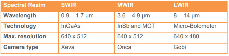
Table 1. Image sensors and cameras offered by Xenics cover the entire infrared realm at high resolution and sensitivity. Source: Xenics.
Midrange IR Metal Detector
Whereas art examination using the short wave infrared has become a standard technique, intensive R&D work is directed at new procedures in the midrange infrared (MWIR) covering wavelengths from 3 to 5 μm.
An examination that was recently carried out by an Italian research team in the Dome at Monza, Italy, shall serve as an example of this new procedure [3]. The researchers have developed a non-invasive method called Thermical Quasi Reflectography. TQR can be applied in-situ. Midrange infrared radiation of pigments usually is captured thermographically. However, TQR uses the reflected MWIR radiation. In this setup, a halogen lamp operated at undervoltage is used as light source and a cooled MWIR camera captures the image.
TQR uncovers features that can not be revealed with SWIR procedures, nor by thermographic methods. This is especially true in the case of investigating wall paintings applied on metal foils as was customary in the late medieval period. Figure 4 shows a section of about 80 x 40 cm of a fresco wall decoration in the Theodelinda chapel of the Monza Dome. It was done by the local Zavattari family between 1440 und 1446.
The image in Figure 4 was taken in the visible spectrum, followed by SWIR capture and finally as TQR image in the MWIR realm. It clearly shows that the gold and sliver foils reflect the light in the MWIR significantly stronger than in the SWIR. In addition, the MWIR capture differentiates much better the pigments barely separarable in the SWIR. According to Table 1 there are several powerful cameras available for this purpose.
Following these ideas, it will be very interesting to observe the art examination techniques as they might develop in the LWIR realm from 8 to 14 μm.
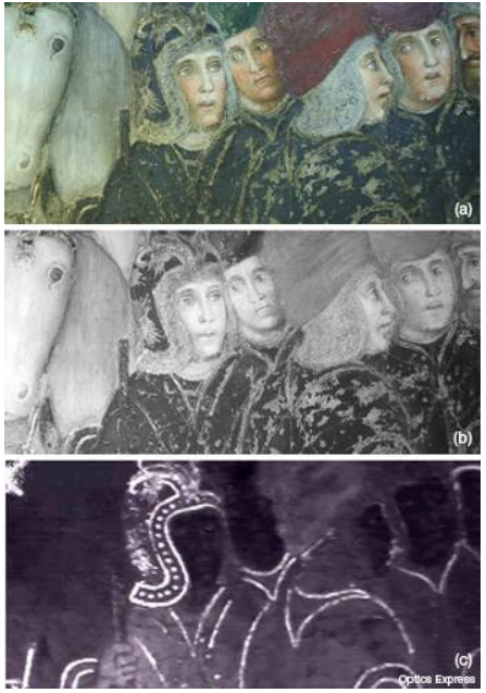
Figure 4. TQR examination carried out in the MWIR realm unveils the use of gold and silver foils in medieval wall frescos. Source: [3].
Terahertz: Another Alternative
Promising analysis methods are developing at a fast pace, also in the neighbouring spectral area of Terahertz frequencies (Figure 1). Now that sufficiently powered radiation sources have become available, the search for appropriate application areas is on – also in the area of art inspection. Since Terahertz frequencies can penetrate much deeper into objects under test, they are well suited for the depth analysis of paint layers on substrates such as canvas, ceramics, and walls.
A typical example of a Terahertz examination is given in Figure 5. It shows how a text is read through a book cover. This is relevant in case an ancient manuscript or document inside a book can not be read because any attempt to open it would irrevocably damage it. This leaves no other choice than reading – or rather reconstructing – the pages through the cover or envelope by applying Terahertz radiation.
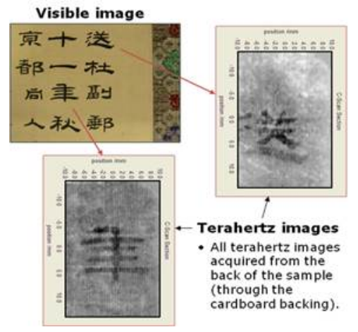
Figure 5. Terahertz capture of a Chinese text through the closed cover of a book. Source: Teraview.
Conclusion
The continuing development of art examination techniques and tools based on radiation methods in the IR to Terahertz ranges and their availability to museums and research groups will further a viable conservation of our common cultural heritage for future generations.
Documentation
Application Note


Related content

Munich.
FROM Nov 10th 2026 TO Nov 13th 2026
SEMICON EUROPA 2026
Join Exosens at SEMICON EUROPA 2026 2026 in Munich

Edinburgh.
FROM Sep 14th 2026 TO Sep 17th 2026
SPIE Sensors + Imaging 2026
Join Exosens at SPIE Sensors + Imaging 2026 Edinburgh, United Kingdom

Jul 07th 2026
Multifocal Femtosecond Laser Writing
Crosstalk Limits in Multifocal Parallel Femtosecond Direct Laser Writing: Decoherence Strategies and Quantitative Validation of Nanostructure Optical Phase

Jun 29th 2026
Exosens secures contract with BROLIS
Exosens secures major long-term contract with BROLIS to supply 17000 high-performance image intensifier tubes for the Czech Armed Forces

Aug 20th 2021
Visit our booth #514 during OPTIFAB

Jun 25th 2026
Strengthening Defense & Security Innovation
Exosens secures €140 million in EIB financing to foster innovation in Europe’s defense and security industry