ABSTRACT
Researchers at the Erasmus MC University Medical Center and at RiverD International B.V., Rotterdam, the Netherlands, have developed a novel, cutting-edge method of applying shortwave-infrared Raman spectroscopy at a wavelength above 1 µm to the examination of darkly pigmented human tissue, thereby greatly subduing the spectral disturbances by fluorescence effects in the visible realm. This will substantially further and accelerate the diagnosis of melanoma. Key to this unique achievement is an image capture technology developed by Xenics, based in Leuven, Belgium. Their LN2-cooled Cougar-640 camera features an extremely low-noise (<20e- typically) InGaAs area array detector with 640 x 512 pixels and a special nondestructive readout scheme called “Read While Integrate.”
The Starting Point: InGaAs instead of CCD
The low read-out noise obtained by the Xenics detector technology can be effectively lowered further as shown in this article. The detector is well suited for all Raman measurements that may or must be shifted to the shortwave infrared (SWIR) spectral range. The camera and its focal-plane detector array were designed by Xenics for scientific and high-end industrial image capture applications. To be introduced to everyday medical practice and to meet its prevailing cost constraints, an array with fewer pixels may be sufficient for medical Raman spectroscopy applications, including the melanoma diagnostics described here.
The Xenics Cougar-640 was introduced in 2012 as a high-performance camera for extreme low- light-level imaging applications in the shortwave infrared realm. By means of its LN2 cooling system it reduces dark current to extremely low levels. The spectral response covers the wavelength range from 0.9 to 1.55 μm (at ~77K sensor temperature). The camera incorporates an in-house designed InGaAs focal plane array detector (XFPA-1.7-640-LN2) intended for fluorescence imaging and photon emission failure analysis applications in the 0.9 to 1.55 µm range.
The detector array features 640 x 512 pixels at a pixel pitch of 20μm complemented by a 24-bit ADC in the camera. It is optimized for operation at 77K with liquid nitrogen (LN2) cooling. The topology used is based on an SFD (source follower per detector) read-out scheme to achieve ultra-low noise levels of 20 e- (at 77K, with correlated double sampling to cancel the read noise related to capacitor offset). Each pixel features a full-well capacity of about 480 000 electrons and a conversion gain of 2.17 μV/e‐. The dark current is typically lower than 20 e-/sec/pixel, at 77K sensor temperature and with a target emissivity of 5% and target temperature at 300K.
With a cooled target (~77K), even lower dark current values can be achieved. The extremely low dark current allows integration times of several hours. A non-destructive read-out mode (also called Read While Integrate) simplifies its operation when long integration times are required. Read-out mode is either Integrate Then Read (can be used with cooled or uncooled sensor) or Read While Integrate (main use is for very long exposures at 77K sensor temperature).
In Read While Integrate (RWI) mode at 77K, the Cougar is suited for extremely low-light-level applications. It is intended to offer the lowest noise and highest sensitivity for industrial applications such as photoluminescence measurements in semiconductor manufacturing and failure analysis, astronomy and general Raman spectroscopy applications. Also, demanding applications in the food processing industry, such as inspecting incoming deliveries of fruit and vegetables, are appropriate field of use.
The camera setup consists of two modules: a Dewar (178 x 93 x 207 mm³, LxWxH) containing the LN2-cooled InGaAs sensor in vacuum, and a separate housing for the control and read-out circuitry (40 x 100 x 130 mm³). The camera interface is provided via standard CameraLink for ease of integration in measurement systems (Figure 1).
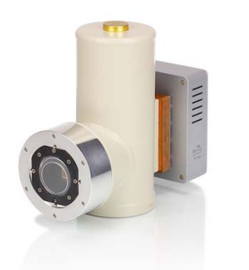
Figure 1. The Xenics Cougar camera containing the InGaAs focal plane detector suited for image capture in the shortwave infrared. Source: Xenics.
Progress in Raman Spectroscopy
“Raman spectroscopy is a very versatile technique to look at anything that you can shine light on,” says Gerwin J. Puppels, one of the six researchers at the Erasmus MC University Medical Center Rotterdam who collaborated with RiverD International B.V. in an extensive medical Raman spectroscopy project funded by the Netherlands Ministry of Economic Affairs. The results were published in April 2015 in the Journal of Raman Spectroscopy [1].
Raman spectroscopy is a powerful and sensitive, non-destructive and non-contact analysis quantitative method delivering high detail. It is based on the measurement of the inelastic scattering of light from the molecules of the investigated material when illuminated by monochromatic light from an appropriate source, usually a laser, at low light levels, basically on photon counting.
The photons of the illuminating light source are absorbed by the sample and re-emitted with a slightly – up or down – shifted frequency. This is the basic Raman phenomenon, discovered by and named after the Indian Scientist Chandrasekhara Venkata Raman in 1928, who was subsequently honored with the Nobel Price in 1930. The downshifted frequency, used in the application of the Raman effect, is called the Stokes frequency; its upwards shifted counterpart is called “Anti Stokes.” Stokes and Anti-Stokes frequency parts are just a minuscule fraction of just 0.001% of the total light reflected by the sample due to elastic scattering, called Rayleigh scattering.
Separating the desired Stokes frequency part from the extremely energetic Rayleigh scattering requires an elaborate setup of apertures, filters, multi-spectroscopic and tuning devices to block the intense stray light primarily caused by the spectrometer’s grating devices. To overcome these basic difficulties, several different methods were conceived and developed for sample illumination and detection, such as stimulated irradiation, coherent anti-Stokes or non-linear stimulation, surface-enhanced Raman spectroscopy using signal emission from metallic surfaces, and others. Nevertheless, despite these adverse conditions, Raman spectroscopy has successfully evolved its uses for scientific and industrial purposes over the last ten to twenty years.
In the course of the historical development of Raman spectroscopy the detector of choice has usually been a Silicon-based CCD sensor operating in the visible realm of the spectrum. Its disadvantage is that its usable responsivity range peters out at and above a wavelength of 1 µm. This turns out to be a serious limitation in the intended medical applications, especially when it comes to investigate and characterize starkly pigmented bio-samples such as human tissue. The reason: in the visible spectrum these tissue samples tend to emit a disturbingly strong fluorescence, which severely impacts the obtained Raman spectra.
There have been numerous attempts to mitigate the analytical degradation by fluorescence effects. Among of those methods are time-gated detection, photo bleaching, confocal signal detection, surface enhanced Raman spectroscopy (SERS) and resonance Raman (RR) scattering (see original paper [1] for detailed author references). All of these solutions have not led to a suitable setup for in-vivo Raman spectroscopy of pigmented tissues.
Another method tried out has been Fourier-transform (FT) Raman spectroscopy. It will yield usable spectra of pigmented skin lesions as indicated in the research literature (see reference). FT Raman spectroscopy proves that tissue fluorescence can be successfully sidetracked by deploying a longer laser excitation wavelength (such as 1064 nm). The drawback, however, is that FT-Raman spectroscopy is based on the multiplexing of single-channel analysis. This leads to signal integration times, which can be longer by several orders of magnitude than with multi- channel Raman spectroscopy.
Raman Spectroscopy in the SWIR
Therefore the six researchers at the Erasmus Medical Center decided to follow a different path in their medical Raman spectroscopy project. They too used a higher frequency range of laser light excitation to avoid the generation of fluorescence in pigmented tissue. But in this process they had to overcome the fact that until recently there was no camera or detector available for the wavelength range above 1 µm that would be comparable to a CCD camera in terms of read noise, which was and is the preferred tool for Raman spectroscopy – if only in the visible spectrum.
Generally, the main limitation of Raman spectroscopy is the noise floor due to the extremely weak intensity of the Raman signal. This limitation is aggravated if the detector adds additional readout noise. In a CCD camera this tends to be in the neighborhood of two or three electrons. In contrast, InGaAs cameras, at least until recently, delivered a readout noise of up to several hundred of electrons, a value not suited for being used in this context.
In Raman spectroscopy, Puppels says, the measurement basically consists of devising the most appropriate measurement setup for capturing all available photons. “If you observe a signal, say 10,000 photons, the shot noise on the signal is 100 photons. This would give you a signal-to- noise ratio of 100. Unfortunately, the read-out noise of the detector will typically add an additional several hundreds of electrons, and the Raman measurement is no longer shot noise limited. That puts you in a very bad position – especially when encountering weak signals consisting of just 10,000 photons.” This condition limits the application of Raman spectroscopy especially when looking for novel medical uses such as melanoma diagnostics.
The basic problem in medical research and diagnostics when considering Raman spectroscopy as an everyday tool is the fact that a melanoma is typically darkly pigmented. This pigment induces strong fluorescence effects in the spectra observed.
Fourier Transform Raman spectroscopy was also considered by the Erasmus MC researchers, but soon rejected. It can deliver satisfying results, even in the presence of pigmented tissue.
However, in this context it is compromised by being a very slow process. It involves a signal integration time of a minute or tens of minutes – just for investigating the results in one single spectrum. Puppels: “It provides nice scientific results but is hardly usable in medical practice.”
Now, with the Cougar camera and its InGaAs detector array available on the market offering a readout noise of less than 20 electrons, the situation has dramatically improved for Raman spectroscopy – in the direction of shifting it to the shortwave infrared realm. Puppels: “This might be a very promising avenue opening up to collect the Raman spectrum in seconds instead of minutes and hours.” The new camera is specified for the wavelength range above 1 µm.
Experimental Setup for Raman Spectroscopy
In the course of their research project at Erasmus MC University Medical Center the team has devised and built a Raman spectroscopy setup, of which the Cougar camera and its InGaAs focal plane detector was a central part. The experimental setup has demonstrated that under these auspices Raman spectroscopy can be successfully used in medical practice delivering high quality HWVN (High Wavenumber) Raman spectra with a low fluorescence background.
To show the feasibility of the wavelength-shifted Raman spectroscopy a complete SWIR multi- channel Raman instrument was constructed (Figure 2).
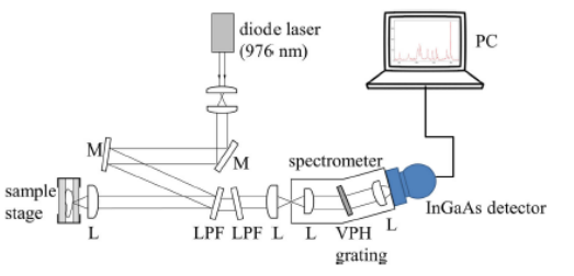
Figure 2. Experimental setup of the SWIR Raman spectroscopy used in the Erasmus MC Medical Research Center Rotterdam research. Source: Journal of Raman Spectroscopy, 2015.
As light source a single-mode continuous-wave diode laser was deployed radiating at a wavelength of 976 nm. It provided an output power of 150 mW (Model R-type from Innovative Photonic Solutions). Considering its scientific and industrial specifications, the Xenics Cougar camera is extremely sensitive at a very low noise level. This makes it an appropriate candidate for medical spectroscopy. “When we investigated the camera,” says Erasmus MC co-researcher Peter J. Caspers, “we found that it listed Raman spectroscopy as one of its applications. But no one had used it for this purpose. The technology is well suited for Raman Spectroscopy and that makes it interesting for our field of medical research.”
Especially the Cougar’s readout scheme “Read While Integrate,” Caspers says, is interesting in that it enables lowering the readout noise by an order of magnitude. The RWI principle was originally implemented in the camera for industrial applications, such as inspecting semiconductor chips for leakage effects. This measurement takes time while the signal builds up. “Our software package does a similar thing to get the Raman signal,” Caspers says. “And because you can read out the signal non-destructively many times before the final result averages the noise over all these readouts we can read it out at a very low effective noise level.”
Read-While-Integrate probes the accumulating photoelectrons in a non-destructive sampling method during integration, without resetting the buffering capacitors. This virtually eliminates the detector’s effective readout noise. RWI is sometimes described as up-the-ramp readout (see reference).
The Cougar detector readout noise (in e-, with CDS) was found to be 22.7 + 5.9 electrons (standard deviation), and dark current (e-/s/pixel) at 69.4 + 4.5 electrons (standard deviation). Note that dark current strongly depends on target temperature and target emissivity. This noise level substantially decreases with more sampling readouts during the integration time. The final results given in Table 1 indicate that the effective readout noise was reduced to the value comparable to cooled slow-scan CCD detectors.
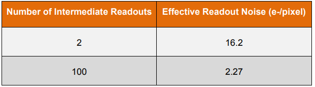
Table 1. Effective readout noise levels obtained with the InGaAs detector of the Cougar camera. Source: Journal of Raman Spectroscopy, 2015.
Since the Cougar camera had never been used for medical spectroscopy, Caspers continues, “some of its characteristics had to be enhanced for our Raman spectroscopy project, for instance its non-linearity properties. This is a part of the software that we developed in addition to the principle that was originally supplied by Xenics.”
Among the specific measures developed to use the camera for the intended shortwave Raman spectroscopy were some algorithms to read and pre-process the raw data that is delivered in the camera’s RWI readout scheme. Also, the camera’s response curve showed a definite progressive non-linear behavior when the accumulated signal exceeded a certain threshold, which necessitated a cutoff threshold for a more linear behavior. An algorithm was devised to correct this non-linear response above the threshold. For this purpose, a first-order polynomial was fitted to the linear range during the first part of the integration period.
Figure 3 shows some tissue samples obtained with the experimental shortwave IR Raman spectroscopy setup.
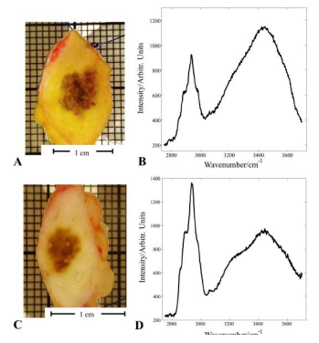
Figure 3. Photographs and Raman spectra obtained with the experimental setup at Erasmus MC of pigmented human tissue indicating a melanoma (A and B), and benign melanocytic tissue (B and C). Laser wavelength: 976nm, exposure time 10s. Source: Journal of Raman Spectroscopy, 2015.
Promising Outlook for Medical Practice
There are still some technical challenges to overcome, the Erasmus MC researchers are cautioning. Raman spectroscopy generally yields a very low signal-to-noise ratio, which puts up some significant hurdles for performing a Raman spectroscopy in the shortwave infrared to circumvent the fluorescence effects in the visible realm produced by pigmented tissue – not the least of which are cost considerations.
Caspers: “One of the many things that we appreciate in the camera is its non-destructive readout. So we can sample it as often as we want – and thereby significantly reduce the effective readout noise. This way we went from about 20 electrons to about two electrons, effectively.” This is the same level as with current CCD cameras. But we are now getting promising results in a new wavelength realm.”
References
[1] Santos, Inês P., Caspers, Peter J., Bakker Schut, Tom, van Doorn, Remco, Koljenovićc, Senada and Puppels, Gerwin J.: Implementation of a novel low-noise InGaAs detector enabling rapid near-infrared multichannel Raman spectroscopy of pigmented biological samples. Journal of Raman Spectroscopy, 2015 (wileyonlinelibrary.com) DOI 10.1002/jrs.4714. E-mail: p.caspers@erasmusmc.nl
Documentation
Application Note


Related content

Munich.
FROM Nov 10th 2026 TO Nov 13th 2026
SEMICON EUROPA 2026
Join Exosens at SEMICON EUROPA 2026 2026 in Munich

Edinburgh.
FROM Sep 14th 2026 TO Sep 17th 2026
SPIE Sensors + Imaging 2026
Join Exosens at SPIE Sensors + Imaging 2026 Edinburgh, United Kingdom

Jul 07th 2026
Multifocal Femtosecond Laser Writing
Crosstalk Limits in Multifocal Parallel Femtosecond Direct Laser Writing: Decoherence Strategies and Quantitative Validation of Nanostructure Optical Phase

Jun 29th 2026
Exosens secures contract with BROLIS
Exosens secures major long-term contract with BROLIS to supply 17000 high-performance image intensifier tubes for the Czech Armed Forces

Aug 20th 2021
Visit our booth #514 during OPTIFAB

Jun 25th 2026
Strengthening Defense & Security Innovation
Exosens secures €140 million in EIB financing to foster innovation in Europe’s defense and security industry